 Home
HomeIntro
In this section
| • | Find out what makes a good specimen |
| • | Look at still fish in different ways |
| • | See how motion adds to your observation |
| • | Look at different ways to look inside |
 Image source details
Visualizing fishes
"Seeing is believing" and "a picture is worth a thousand words" - these phrases capture the importance we place on visual information. We use natural and technology-enhanced methods to see - to observe and show anatomy, to view patterns of activity over space and time, and to explain and convince.
Observation is an essential activity of science and nearly everything we do. What does a particular species of fish look like? How are the parts of a fish eye configured? What does a parrotfish do all day? Where can we hunt for tuna? These questions require observations and visualizations to answer.
Observation is beyond vision - In this section, we present an introduction to the ways researchers observe fish and how those observations are shared with others.
Some methods of looking at fishes such as photography, microscopy, and scientific illustration are still very useful although developed long ago. Many techniques involve detection of electromagnetic radiation beyond visible light.
The cutting-edge technologies allow us to extend our range of vision - to see things that are even smaller, farther away, hidden, distributed over a large area, or fleeting in time. Magnetic resonance imaging (MRI) is one of the newest technologies we highlight. MRI lets us look at internal organs in three dimensions without slicing any fish open. For rare specimens, keeping them intact is an advantage. Using stored 3-D digital data, it can be viewed using special software allowing us to produce images on-the-fly, as if we were looking at a real sample and changing our point of view at will.
Other techniques involve visualizing individuals and groups of fishes where they live. Since sound travels through water better than light, some observation rely on sound rather than radiation.
Enjoy the view!
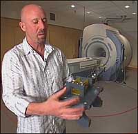
Center for Functional Magnetic Resonance Imaging
Photographer - TBA
The Keck Center at UCSD's School of Medicine is a state-of-the-art imaging facility for basic and translational research.
Recognizing the need to provide dedicated research MRI systems for San Diego's large and diverse neuroscience community,the center was established in 2002 in collaboration with the Salk Institute for Biological Studies. Today, imaging scientists from UCSD and affiliated institutions are engaged in a wide range of studies that make use of the full spectrum of MRI imaging methods to study the metabolic, neural,vascular and anatomical substrate of normal and disordered brain function.
In addition, the Center is dedicated to fostering collaborations across traditional disciplines, building on UCSD's neuroimaging expertise to create a unique and interdisciplinary environment for scientists from the departments of bioengineering, orthopedics, Scripps Institution of Oceanography and others. More information is available at http://cfmri.ucsd.edu/.